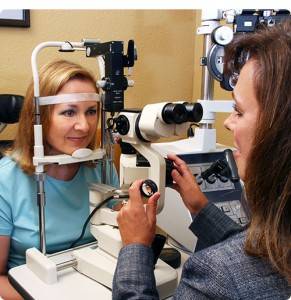
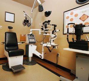
Your vision is your window to the world!
 But without proper eye care, you might not know how much you’re missing. Conditions like Macular Degeneration, Glaucoma and Cataracts can occur silently and may cause permanent damage. Protect and perfect your vision by scheduling annual eye exam with Cove Eyecare.
What to expect on your first visit:
We want to make your experience as comfortable, personalized and on time as possible. Please arrive 15 minutes before your appointment. Be certain to bring a list of all your current medications and eye drops as well as insurance information, Military ID and health insurance cards. Our Patient Intake Forms can be filled out online. Please do so in advance of your appointment.
- Basic Examination
- Pretest
- Examination
- Comprehensive Examination
- Contact Lens Fitting
- Urgent Care Visits
Advanced Diagnostic Testing
- Digital Fundus Photography
- Visual Field Test
- Ocular Coherence Tomography (OCT)
- OCT 3D Visualizations
- Corneal Pachymetry
- Anterior Chamber Angle Assessment
What Does A Basic Eye Exam Include?
A basic examination includes the following tests and procedures:
Pretest:
Entering acuities, lensometry of current vision prescription, screening visual fields, stereopsis, fusion, muscle balance, color vision, autorefraction, autokeratometry and screening digital retinal photos.
Examination:
 Examination includes a review of your medical, family and ocular history and discussion of your current visual concerns and symptoms. Testing of the pupillary reflexes, manifest refraction at distance and near to determine spectacle prescription. Best corrected acuities, measurement of intraocular pressure, evaluation of the anterior structures of the eye and adnexa with biomiscroscopy, review of retinal photos and retinal evaluation with direct and indirect ophthalmoscopy. Test results are reviewed, ocular health and refractive status is discussed as well as any ocular concerns are addressed. Treatment options are reviewed and recommendations are made for the best correction of vision, a spectacle lens prescription is finalized and additional diagnostic testing is scheduled if indicated.
Comprehensive Eye Exams
The comprehensive examination includes pupillary dilation and is recommended for individuals over the age of 50, high myopes, diabetics, hypertensives, individuals who have been diagnosed with cataracts, glaucoma or macular degeneration and anyone who is experiencing flashes of light or floaters.
In addition to all of the procedures in a basic examination, central retina is evaluated via biomicroscopy, peripheral retinal evaluation is performed with a binocular indirect ophthalmoscope, digital retinal photos are performed after dilation, and a Cirrus HD-OCT is used to perform scans of the macula, optic nerve, anterior chamber and cornea.
Contact Lens Fitting:
Performed upon patient’s request during a Basic or Comprehensive Exam. A contact lens history is taken, wear and care habits are reviewed, and recommendations are made as to new products to promote better corneal health, reduce redness, to mitigate dryness or loss of near focus. Contact lens fittings include free trial lenses, a care kit, instruction on insertion and removal if needed, and follow up visits until the contact lens prescription is finalized.
Urgent Care Visits
Urgent care visits involve a history and timeline of urgent eye conditions or injuries, appropriate examination which may include pupillary dilation if indicated, treatment and follow up.
Advanced Diagnostic Testing During Your Eye Exam
![fort hood eye exam]() Digital Fundus Photography
Digital Fundus Photography
Digital Fundus Photography is specialized medical imaging used to take pictures of the structures located at the back of the eye, including the retina. It produces a series of photos that are helpful for diagnosing, documenting, and monitoring certain eye conditions. Fundus photography is a short, painless procedure.
Visual Field Test
Visual Field Test is used to map and monitor the sensitivity of the field of vision. You will be seated with your chin in the the Humphrey 450i, looking into a concave dome with a target in the center. The eye that is not being tested is covered. A point of light is presented while you concentrate on a central target straight ahead, and you are asked to press a button when you see a point of light. Your responses are analyzed statistically, compared to normal responses and archived for comparison to future tests. If a test result is abnormal, the test may be repeated to help verify the first test was correct or to confirm a visual field defect. Visual filed analysis is critical in detecting and managing numerous ocular pathologies such as glaucoma, optic nerve disease, vision loss associated with stroke and aging changes of the upper eyelids, as well as monitoring individuals who are taking high risk medications with serious adverse visual side effects. The test takes 5 to 8 minutes per eye and it is important to be alert, focused and making your best effort during the test.
Ocular Coherence Tomography (OCT)
Ocular Coherence Tomography (OCT) is a state-of-the-art imaging method used to evaluate the eye for subtle changes when early detection and treatment can be the most beneficial in preventing vision loss.
It is the most advanced tool available to help manage conditions such as glaucoma, age-related macular degeneration, diabetic retinopathy, epiretinal membranes, macular swelling, macular holes, cystoid macular edema, central serous retinopathy, and optic nerve damage. It is non-invasive, quick, painless.
![eye exam technology in fort hood]() Cirrus HD-OCT
Cirrus HD-OCT
Our Cirrus HD-OCT uses scanning lasers and sophisticated technology to paint 3D images of the most critical structures of the eye, analyze the results statistically and compares the results to a normative, FDA approved database. This allows for precise evaluation and is invaluable in detecting even the slightest change over time.
Corneal Pachymetry
Corneal Pachymetry is an important element in the management of patients diagnosed with glaucoma, as well as those at high risk for developing glaucoma (i.e., glaucoma suspect). Pachymetry can also be helpful in the diagnosis and measurement of corneal dystrophies such as Fuch’s endothelial dystrophy and conditions such as keratocunus. Corneal pachymetry is the measurement of corneal thickness using ultrasound. Ultrasound energy is emitted from the probe tip acting as both the transmitter and receiver. It is a painless, rapid and non-invasive measurement.
Anterior Chamber Angle Assessment
Anterior Chamber Angle Assessment / OCT is a new method using the OCT to provide high resolution images of the angle formed by the cornea and iris. This anatomic location is critical in evaluating glaucoma and detecting eyes at risk for angle closure.


 Digital Fundus Photography
 Cirrus HD-OCT

